Have You Noticed Small White Spots on Your Arms or Legs? Here Is Exactly What They Are
Few things are as quietly unsettling as discovering a constellation of small, pale spots on your skin that were not there before. You might notice them while applying lotion after a shower, catching a glimpse of your forearm in the sunlight, or simply looking down at your legs. They do not itch, they do not hurt, and they seem to have appeared out of nowhere. For many people, the immediate reaction is a quiet spiral of worry: Is this dangerous? Is it a sign of something serious? Will it spread?
The good news is that the overwhelming majority of small white spots on the arms and legs are entirely harmless. In fact, the most common cause is a benign, incredibly widespread condition that affects the vast majority of people as they age, particularly those who have spent time in the sun. However, “harmless” does not mean “identical.” Several different skin conditions can produce white spots, and they each have distinct characteristics, underlying causes, and management strategies. What looks like a simple sun spot to one person may be a fungal infection or an autoimmune condition in another.
Understanding the difference is essential, not only for peace of mind but also for choosing the right approach to care. Some spots require nothing more than a good sunscreen and a shift in perspective, while others benefit from targeted medical treatment to prevent progression or relieve symptoms. This comprehensive guide will walk you through the most common causes of small white spots on the arms and legs, explain the science behind why they form, detail the available treatment options, and clarify the warning signs that mean it is time to see a dermatologist.
Understanding Skin Pigmentation: The Foundation of White Spots
To understand why white spots appear, it helps to understand how skin gets its color in the first place. Human skin contains specialized cells called melanocytes, which are located in the basal layer of the epidermis, the outermost layer of the skin. These cells produce melanin, the pigment responsible for the color of your skin, hair, and eyes. Melanin also serves a critical protective function: it absorbs ultraviolet (UV) radiation from the sun, shielding the deeper layers of the skin from DNA damage that can lead to cancer.
When something disrupts the production of melanin or interferes with the function of melanocytes, the affected area loses its color. This condition is known broadly as hypopigmentation. The white spots you see on your arms or legs are, in essence, localized areas where the skin has lost some or all of its pigment. The specific reason for this loss—whether it is sun damage, a fungal overgrowth, an autoimmune reaction, or simply the wear and tear of life—determines the exact appearance of the spots, their location, and the best way to manage them.
Idiopathic Guttate Hypomelanosis: The Most Common Cause
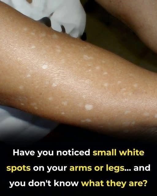
If you are an adult, particularly one over the age of 40, and you have noticed small, flat, porcelain-white spots on your forearms or shins, the most likely explanation is idiopathic guttate hypomelanosis (IGH). Despite its intimidating name, IGH is a benign, noncancerous condition that is incredibly common. The name itself is descriptive: idiopathic means the cause is unknown, guttate means “resembling drops,” and hypomelanosis means a lack of melanin.
What Does IGH Look Like?
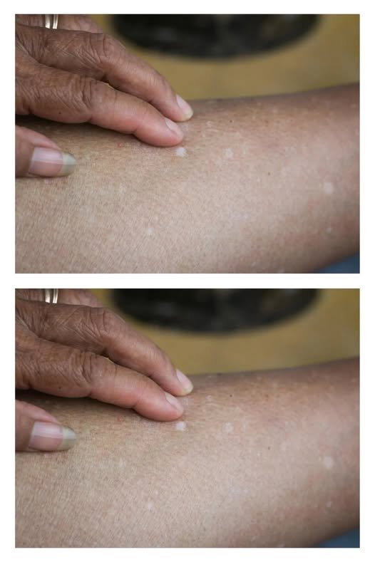
IGH presents as multiple small, sharply defined white or off-white spots. They are typically round or oval and measure between 2 and 5 millimeters in diameter, though some can be slightly larger. The spots are usually smooth to the touch, with no scaling or crusting, and they do not itch or cause any physical discomfort. They are most commonly found on sun-exposed areas, particularly the forearms and shins, which is precisely where the image in question shows them. Less frequently, they can appear on the face, neck, or trunk. The number of spots tends to increase with age; some individuals may have just a few, while others may have over a hundred.
Who Gets IGH and Why?
IGH affects people of all skin colors, but it is most frequently observed in fair-skinned individuals. While it can appear in young adults, its prevalence rises sharply with age. Research indicates that up to 87% of individuals aged 40 and older have at least one lesion, and up to 80% of people over 70 are affected.
The exact cause of IGH remains unknown, but the scientific consensus points to a combination of factors. Cumulative, chronic sun exposure is considered a central contributor, as the spots appear predominantly on sun-exposed areas. Aging is another key factor, as the condition becomes increasingly common in older populations. Some researchers also implicate genetics and repeated microtrauma to the skin, such as from body scrubs or friction. In essence, IGH is best understood as a marker of the skin’s natural aging process and the cumulative effects of environmental exposure.
Is IGH Dangerous?
No. IGH is completely benign. It does not predispose a person to skin cancer, nor is it a sign of an underlying systemic disease. The primary concern is cosmetic. Because the spots lack melanin, they do not tan. This means that when the surrounding skin darkens in the sun, the white spots stand out even more prominently, which can be distressing for some individuals.
Vitiligo: An Autoimmune Pigment Disorder
While IGH is a cosmetic issue related to sun damage and aging, vitiligo is a chronic autoimmune condition with more significant implications for skin health. Vitiligo occurs when the body’s immune system mistakenly targets and destroys the melanocytes in the skin, leading to progressive depigmentation.
Recognizing Vitiligo
The white patches of vitiligo are distinctly different from the small, scattered dots of IGH. Vitiligo typically presents as larger, well-defined patches of completely white or chalky skin. These patches often have a symmetrical distribution and can appear on both sides of the body. While vitiligo can affect the arms and legs, it frequently begins around the mouth, eyes, nose, hands, feet, elbows, and knees. Unlike IGH, vitiligo patches can enlarge and spread over time if left untreated. The depigmented skin is highly vulnerable to severe sunburn because it lacks the protective melanin barrier.
How Vitiligo Is Diagnosed and Treated
A dermatologist can usually diagnose vitiligo through a clinical examination. A Wood’s lamp—a special handheld device that emits ultraviolet light—is often used to enhance the appearance of depigmented patches and reveal any subtle involvement that may not be visible under normal lighting. Skin scrapings may be performed to rule out fungal infections.
Treatment for vitiligo aims to restore pigment, stabilize the condition, and protect the skin. First-line therapies include topical corticosteroids, which help suppress the autoimmune response, and calcineurin inhibitors such as tacrolimus and pimecrolimus. For more extensive or stubborn cases, narrowband ultraviolet B (UVB) phototherapy is often the treatment of choice. In recent years, newer topical treatments specifically indicated for repigmentation have been approved, offering additional options for patients.
Tinea Versicolor: A Fungal Cause of White Spots
Not all white spots are related to pigment cells. Tinea versicolor (also known as pityriasis versicolor) is a common fungal infection caused by an overgrowth of Malassezia yeast, which naturally lives on human skin.
The Fungal Mechanism
The Malassezia yeast produces a substance that interferes with melanin production, resulting in areas of skin that are lighter than the surrounding tissue. These patches may also prevent tanning, making them more noticeable after sun exposure. Tinea versicolor most commonly affects areas rich in sebaceous (oil) glands, such as the chest, back, and upper arms. However, it can also occur on the legs. Unlike IGH, the patches of tinea versicolor often have a fine, scaly surface and may be mildly itchy, especially in warm, humid conditions.
Diagnosis and Antifungal Treatment
Diagnosis is typically straightforward. A dermatologist may use a Wood’s lamp, under which tinea versicolor may fluoresce a characteristic golden-yellow color. A skin scraping examined under a microscope can confirm the presence of fungal elements. Treatment involves the use of antifungal medications. For localized cases, topical antifungal shampoos, lotions, or creams are usually sufficient. Common options include selenium sulfide shampoo, ketoconazole cream or shampoo, or pyrithione zinc soap. For extensive or recurrent cases, oral antifungals may be prescribed. It is important to note that even after the fungus is eliminated, the white patches may take months to regain their normal pigmentation.
Pityriasis Alba: The Childhood Condition
Pityriasis alba is a common condition that primarily affects children and young adults, though it can occur at any age. It causes round or oval patches of lighter skin, often with a slightly raised, scaly border.
What Causes Pityriasis Alba?
The exact cause is not fully understood, but it is thought to be related to sun exposure and dry skin, and it is often associated with a history of eczema or atopic dermatitis. The lightening is thought to result from low-grade inflammation that disrupts normal melanin production. These patches are most often seen on the face, upper arms, and trunk.
Management and Prognosis
Pityriasis alba usually improves on its own over time, although it may take months or even years for the skin color to return to normal. Moisturizing the skin regularly with a fragrance-free cream or petroleum jelly can help relieve dryness and support the skin barrier. If the patches are itchy or irritated, a doctor may recommend a mild topical steroid cream. Sun protection is also important, as sun exposure can make the contrast between the light patches and the surrounding tanned skin more noticeable.
Post-Inflammatory Hypopigmentation: White Spots After Skin Injury
Sometimes, white spots appear in the exact location where the skin was previously inflamed or injured. This is known as post-inflammatory hypopigmentation. It occurs when inflammation disrupts the normal function of melanocytes, leading to a temporary decrease in pigment production.
Common Triggers
Post-inflammatory hypopigmentation can follow various skin conditions, including eczema, psoriasis, burns, insect bites, or even minor trauma such as scratches. It is particularly common and noticeable in people with darker skin tones. The spots are usually flat and correspond to the shape and location of the original inflammation. They are not typically scaly or itchy unless the underlying inflammatory condition is still active.
Management and Prognosis
The most important step in managing post-inflammatory hypopigmentation is to treat the underlying inflammatory condition. Once the inflammation is controlled, the skin often repigments on its own over time, although this can take several months. To aid the process and make the spots less noticeable, strict sun protection is essential. Sunscreen prevents the surrounding skin from tanning, which would otherwise accentuate the contrast between the normal and hypopigmented areas.
Milia: Tiny White Bumps
While the spots discussed above are flat, milia are tiny, raised, pearly-white bumps that develop when keratin, a protein in skin cells, becomes trapped just beneath the surface of the skin. They are most common on the face, particularly around the eyes and cheeks, but they can also occur on the arms, legs, and trunk.
Milia are harmless and painless. Unlike the flat spots of IGH or vitiligo, milia feel like tiny pearls under the skin. They often resolve on their own, but a dermatologist can safely extract them if they are bothersome. For multiple milia, topical retinoids or procedures such as electrodesiccation may be used.
How to Tell the Difference: A Comparison of Key Features
The visual similarities between these conditions can make self-diagnosis difficult. The following table summarizes the defining characteristics of the most common causes of white spots on the arms and legs.
Condition
Typical Appearance
Common Locations
Sensation
Cause
Idiopathic Guttate Hypomelanosis (IGH)
Small (2-5mm), flat, sharply defined porcelain-white dots
Forearms, shins, sun-exposed areas
Asymptomatic
Sun damage, aging, genetics, microtrauma
Vitiligo
Larger, well-defined patches of chalky-white skin
Often symmetrical: around mouth, eyes, hands, elbows, knees
Asymptomatic; skin is sun-sensitive
Autoimmune destruction of melanocytes
Tinea Versicolor
Scaly patches, lighter or darker than surrounding skin
Chest, back, upper arms; can spread
May be mildly itchy
Overgrowth of Malassezia yeast
Pityriasis Alba
Round or oval, slightly scaly, lighter patches
Face, upper arms, trunk
Mildly itchy or asymptomatic
Associated with eczema and dry skin
Post-Inflammatory Hypopigmentation
Flat, pale areas matching the shape of previous injury
Anywhere the skin was previously inflamed
Asymptomatic
Inflammation disrupting melanin production
Milia
Tiny, raised, pearly-white bumps
Eyelids, cheeks, forehead, arms, legs
Asymptomatic
Trapped keratin
Diagnosis: How a Dermatologist Identifies White Spots
Because several conditions can look similar, a proper diagnosis is essential. A dermatologist can typically diagnose the cause through a simple visual examination. They may use a Wood’s lamp to enhance the appearance of depigmented patches and help distinguish between conditions like vitiligo and tinea versicolor. In some cases, a skin scraping may be performed to check for fungal elements, or a small sample of skin may be taken for a biopsy if the diagnosis is uncertain.
Treatment Options: From Observation to Intervention
For many of the conditions described, treatment is not medically necessary, but it can improve appearance and address any underlying issues.
For IGH
IGH does not require treatment. However, if the appearance is bothersome, several options exist. Topical retinoids (like tretinoin) or topical corticosteroids may be prescribed, though results are variable. Cryotherapy (freezing with liquid nitrogen) is a low-cost, minimally invasive procedure that has been shown to be effective for IGH. Laser therapies, including fractional lasers and excimer lasers, and chemical peels or dermabrasion are also used to reduce the visibility of the spots and improve skin tone. The effectiveness of each treatment varies, and it is important to discuss the risks and benefits with a dermatologist.
For Vitiligo
Treatment is focused on halting the autoimmune attack and stimulating repigmentation. This may involve topical corticosteroids, calcineurin inhibitors, phototherapy, or newer agents specifically approved for repigmentation.
For Tinea Versicolor
Antifungal treatments, available as topical shampoos, creams, or oral medications, will clear the infection. However, the white patches may take months to regain their normal pigmentation after the fungus is gone.
For Pityriasis Alba and Post-Inflammatory Hypopigmentation
The primary strategy is to treat the underlying inflammatory condition, moisturize the skin, and practice strict sun protection to allow the skin to repigment naturally.
The Crucial Role of Sun Protection
Regardless of the specific cause, daily application of a broad-spectrum sunscreen with an SPF of 30 or higher is a universal recommendation for anyone concerned about white spots. Sun exposure can worsen the appearance of IGH, trigger flares of vitiligo, and make post-inflammatory hypopigmentation more noticeable by darkening the surrounding skin. Protecting your skin from the sun also reduces your overall risk of skin cancer, a benefit that far outweighs any cosmetic concern.
When to See a Dermatologist
While most white spots are harmless, certain signs warrant a professional evaluation. You should schedule an appointment with a dermatologist if you notice any of the following:
Rapid Spread: The spots are new, increasing in number, or spreading quickly.
Sharp, Complete Depigmentation: The patches are completely white (chalky) and sharply outlined, which may suggest vitiligo.
Associated Symptoms: The spots are accompanied by itching, redness, scaling, pain, or changes in hair color.
Involvement of Sensitive Areas: White patches appear on the face, hands, genitals, or around the eyes.
No Improvement: The spots do not improve or resolve after a reasonable period.
Uncertainty: You are unsure of the cause and want an accurate diagnosis.
A dermatologist can provide clarity, rule out serious conditions, and recommend a treatment plan tailored to your skin type and goals.
Conclusion: Understanding Your Skin
Small white spots on the arms and legs are a remarkably common finding, and in the overwhelming majority of cases, they are nothing to worry about. For most people, the spots are simply idiopathic guttate hypomelanosis—benign “white sun spots” that reflect a lifetime of sun exposure and the natural aging of the skin. Other common causes include the fungal infection tinea versicolor, the autoimmune condition vitiligo, and temporary post-inflammatory changes following a skin injury.
The key takeaway is that an accurate diagnosis matters. While self-comparison with online images can provide useful clues, a dermatologist is best positioned to distinguish between conditions that look similar but require very different management strategies. If your white spots are stable, asymptomatic, and limited to sun-exposed areas, they are likely harmless. If they are spreading, changing, or accompanied by other symptoms, a professional evaluation will provide clarity and peace of mind. With the right information and appropriate sun protection, you can better understand and care for the skin you are in.
Frequently Asked Questions
Are small white spots on the arms and legs usually serious?
In the vast majority of cases, no. The most common cause is idiopathic guttate hypomelanosis (IGH), a completely benign condition related to sun exposure and aging. However, other conditions like vitiligo or tinea versicolor can also cause white spots, so a professional diagnosis is helpful if you are concerned.
What is the most common cause of white spots on the legs?
Idiopathic guttate hypomelanosis (IGH) is the most common cause of small white spots on the legs, particularly on the shins. It affects a large percentage of people over the age of 40 and is considered a marker of sun damage.
Can white spots on the skin be a sign of a vitamin deficiency?
Yes, in some cases. Deficiencies in vitamins such as B12, C, D, or folate have been associated with the development of white spots. However, this is less common than other causes. A balanced diet generally prevents such deficiencies.
Do white sun spots (IGH) ever go away?
The spots themselves do not typically resolve on their own. However, they are harmless. Using daily sunscreen can prevent new spots from forming and help protect the skin from further damage. Cosmetic treatments are available for those who wish to reduce their appearance.
How can I tell if my white spots are vitiligo or something else?
Vitiligo typically presents as larger, well-defined patches of completely white skin, often appearing symmetrically on both sides of the body. In contrast, IGH spots are small, scattered dots. A dermatologist can use a Wood’s lamp to distinguish between the two.
Is tinea versicolor contagious?
No, tinea versicolor is not contagious. It is caused by an overgrowth of yeast that naturally lives on everyone’s skin. It is triggered by factors like warm, humid weather and excessive sweating, not by person-to-person contact.
When should I see a doctor about white spots on my skin?
You should see a dermatologist if the spots are spreading rapidly, are completely white and sharply defined, are accompanied by itching or scaling, or appear on sensitive areas like the face or genitals. If you are unsure about the cause, a professional evaluation is always a good idea.