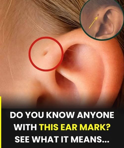
What Is the Tiny Hole in Front of the Ear? The Science Behind Preauricular Pits
Have you ever noticed a small, mysterious opening or dimple located just in front of someone’s ear, near the upper cheek or temple? For many people, this tiny hole is a source of lifelong curiosity, sometimes prompting questions about its origin, its purpose, and whether it is a sign of an underlying health issue. On social media, images of this feature often go viral, accompanied by captions asking, “Do you know anyone with this ear mark? See what it means.” The resulting comment sections are usually filled with a mixture of fascination, personal anecdotes, and wild speculation.
The truth is that this tiny hole is a well-documented medical phenomenon known as a preauricular pit (or preauricular sinus). It is a congenital feature, meaning it is present from birth, and it is far more common than many people realize. While it might look like a piercing, a scar from a childhood injury, or even a bizarre evolutionary leftover, it is actually a fascinating biological quirk rooted in fetal development.
For the vast majority of individuals, a preauricular pit is a completely harmless, asymptomatic curiosity. However, because it is a small tract that dips beneath the surface of the skin, it can occasionally become a site for trapped debris, leading to infections that require medical attention.
This comprehensive guide will explore every aspect of the preauricular pit. We will delve into its embryological origins, examine the genetic and evolutionary theories behind its existence, discuss its prevalence across different populations, outline the potential complications and treatment options, and provide practical advice on how to care for this unique feature. Whether you have one yourself, know someone who does, or are simply curious about this fascinating anatomical variation, this article will provide a clear, evidence-based understanding of the tiny hole in front of the ear.
What Is a Preauricular Pit?
A preauricular pit, medically referred to as a preauricular sinus or preauricular fistula, is a small, blind-ended tract or opening located just anterior to (in front of) the external ear. It is typically found near the upper rim of the ear cartilage, where the front of the ear meets the skin of the face.
Appearance and Anatomy
The opening itself is usually tiny, often measuring just a millimeter or two in diameter. It can look like a small dimple, a deep pore, or a tiny slit. While it appears to be a superficial hole, a preauricular pit is actually the entrance to a narrow, skin-lined tract that extends inward.
The depth and complexity of this tract can vary significantly from person to person. In some individuals, the tract is shallow and ends just beneath the skin. In others, it can branch out into a complex, tree-like structure that extends deep into the surrounding tissue, sometimes even reaching the cartilage of the ear or the parotid gland (the salivary gland located near the jaw).
The Medical Terminology
The term “preauricular” means “in front of the ear.” A “sinus” is a cavity or hollow space within a tissue, while a “fistula” is an abnormal connection between two surfaces. Because the tract is lined with skin cells (epithelium) and does not usually connect to another organ, it is technically a sinus. However, the terms preauricular sinus and preauricular fistula are often used interchangeably in medical literature.
The Embryological Origins: How Does It Form?
To understand why a preauricular pit forms, we must look at the earliest stages of human development in the womb. The ear is one of the most complex structures in the human body, and its formation is a highly intricate process that is vulnerable to minor variations.
The Hillocks of His
During the sixth week of embryonic development, the external ear (the auricle) begins to form from six small swellings of tissue called the auricular hillocks, or the Hillocks of His (named after the German anatomist Wilhelm His, who first described them). These six hillocks are arranged around the first and second pharyngeal arches (sometimes called branchial arches) on either side of the fetal neck.
As the embryo develops, these six separate hillocks grow, merge, and fuse together to form the complex curves and folds of the external ear.
Incomplete Fusion
A preauricular pit occurs when this fusion process is incomplete. If the tissue of the first hillock fails to fuse perfectly with the surrounding tissue, it leaves a small gap or tract behind. As the fetus continues to grow, this tract becomes lined with skin cells and remains as a small opening on the surface of the skin, while the rest of the ear develops normally around it.
Because it is a developmental anomaly, the pit is entirely present at birth, even if it is not noticed until childhood or adulthood. It is not caused by anything the mother did during pregnancy; it is simply a slight variation in the way the fetal tissue folded and fused.
Genetics and Evolutionary Theories
The presence of a preauricular pit is not just a random occurrence; it is often linked to genetics and has sparked interesting debates about human evolution.
Inheritance Patterns
Preauricular pits can occur sporadically (with no family history), but they are frequently inherited. The most common inheritance pattern is autosomal dominant, meaning that a person only needs to inherit one copy of the altered gene from one parent to develop the feature.
If a parent has a preauricular pit, there is a 50% chance they will pass it on to their child. However, the size, shape, and depth of the pit can vary significantly within the same family.
In rare cases, bilateral preauricular pits (pits on both ears) or pits associated with other physical anomalies can be a sign of a genetic condition known as Branchio-Oto-Renal (BOR) syndrome. This is a rare disorder that can affect the development of the branchial arches (which form the neck and ears), the ears (potentially causing hearing loss), and the kidneys. It is important to note that the vast majority of people with preauricular pits do not have BOR syndrome and are otherwise completely healthy.
The Evolutionary Remnant Theory
One of the most popular and persistent myths about the preauricular pit is that it is an “evolutionary leftover” from our distant aquatic ancestors—a remnant of fish gills.
While it is true that the ears and neck develop from the pharyngeal arches (which are homologous to the gill arches in fish), the preauricular pit is not a functioning gill slit. It is simply a structural defect in the fusion of the ear hillocks. However, the term “branchial cleft cyst” or “branchial fistula” is sometimes used for similar anomalies that occur lower down in the neck, which are related to the branchial apparatus. For the preauricular pit specifically, while its location is tied to the embryological structures that evolved into gills, calling it a “gill” is scientifically inaccurate.
Some evolutionary biologists have speculated that the pit may have once served a sensory function, perhaps acting as a pressure sensor or a receptor for low-frequency sounds in our ancient ancestors. However, there is no definitive proof of this, and today, the pit is completely non-functional.
Who Gets Preauricular Pits? Prevalence and Demographics
Preauricular pits are one of the most common congenital anomalies of the head and neck. However, their prevalence varies widely depending on geographic location and ethnicity.
Global Statistics
Worldwide, it is estimated that approximately 1% of the general population has a preauricular pit. This means that in a room of 100 people, there is a good chance at least one person has this feature.
Geographic and Ethnic Variations
The prevalence is not uniform across the globe. The rates are significantly higher in certain populations:
Africa: Prevalence rates in some African countries have been reported to be as high as 4% to 10%.
Asia: Studies in parts of Asia have shown prevalence rates ranging from 2.5% to 5%.
Europe and North America: The rate is generally lower, hovering around 1% or less.
Taiwan: Some studies have shown exceptionally high rates in specific regions, such as Taiwan, where up to 2.5% of schoolchildren have been reported to have the feature.
Unilateral vs. Bilateral
Most people with a preauricular pit have it on only one side (unilateral). Bilateral pits (present on both ears) occur in approximately 25% to 50% of cases. When the pit is bilateral, the likelihood of it being an inherited genetic trait is higher. There is no significant gender difference; males and females are affected equally.
Are Preauricular Pits Dangerous? The Truth About Complications
For the vast majority of individuals, a preauricular pit is a completely benign, asymptomatic cosmetic feature. It causes no pain, does not affect hearing, and requires no medical treatment. However, because the pit is a tract lined with skin cells, it is susceptible to a specific set of complications.
The Risk of Infection
The most common complication associated with a preauricular pit is infection. The small opening can act as a trap for dead skin cells, natural skin oils (sebum), sweat, and dirt. Over time, these substances can accumulate inside the tract, creating an environment where bacteria can thrive and multiply.
When the tract becomes blocked and infected, it can lead to a preauricular abscess or a cyst.
Symptoms of an Infected Preauricular Pit
It is crucial to recognize the signs of an infection so that it can be treated promptly. Symptoms include:
Pain and Tenderness: The area around the pit becomes sensitive to touch.
Redness and Swelling: The skin surrounding the opening becomes inflamed and swollen.
Warmth: The infected area feels hot to the touch.
Discharge: A foul-smelling, pus-like fluid may drain from the opening.
Fever: In more severe cases, the infection can cause a systemic reaction, leading to a fever or general malaise.
If you or a loved one experiences these symptoms, it is essential to consult a healthcare professional. Attempting to squeeze or drain the infection at home can push bacteria deeper into the tissue, worsening the infection and increasing the risk of scarring.
Treatment and Surgical Options
The treatment for a preauricular pit depends entirely on whether it is asymptomatic or prone to recurrent infections.
Asymptomatic Pits
If the pit is not causing any problems, no treatment is necessary. It is best left alone and simply kept clean. Prophylactic surgical removal (removing it before it ever gets infected) is generally not recommended, as the surgery carries its own risks (such as scarring or damage to nearby nerves) and most pits never become infected.
Treatment for Acute Infections
When a pit becomes infected, the first line of treatment is usually antibiotics. A doctor will typically prescribe a course of oral antibiotics to clear the bacterial infection.
If an abscess has formed (a pocket of pus), it may need to be incised and drained by a medical professional. This involves making a small cut in the abscess to allow the pus to escape, which provides immediate pain relief. However, incision and drainage alone do not remove the underlying tract, meaning the infection is likely to recur.
Surgical Excision
For individuals who suffer from recurrent infections, the definitive treatment is surgical excision. This is a minor surgical procedure performed by a general surgeon, otolaryngologist (ENT), or plastic surgeon.
The goal of the surgery is to completely remove the entire sinus tract, not just the visible opening. Because the tract can branch out unpredictably beneath the skin, the surgeon often uses a special dye to trace the pathway, or uses magnifying loupes to ensure every branch is removed.
If the entire tract is not removed, a small remnant can remain, which can lead to a recurrence of the infection. The surgery is usually performed under local or general anesthesia and has a high success rate.
Common Myths and Misconceptions
Because the preauricular pit is a relatively common feature with a slightly unusual appearance, it has attracted several myths over the years.
Myth: It is a sign of high intelligence or a special personality trait. There is no scientific evidence linking the pit to intelligence or personality. It is purely a physical developmental variation.
Myth: It is a piercing hole that closed up. While it looks like a small piercing, it is a congenital tract that formed in the womb, not a healed wound.
Myth: It is a “gill” leftover. As discussed, while it develops from tissues related to the pharyngeal arches, it is not a functional gill or a direct remnant of aquatic breathing mechanisms.
Myth: Everyone with a preauricular pit will eventually get an infection. This is false. The vast majority of people with a pit will never experience an infection and do not require surgery.
Myth: You can “pop” the hole to clean it out. You should never squeeze, pick at, or attempt to force cleaning objects into the pit. This can introduce bacteria and cause trauma, significantly increasing the risk of infection and scarring.
How to Care for a Preauricular Pit
If you or your child has a preauricular pit, caring for it is generally very simple and requires minimal effort.
Gentle Hygiene
The most important step is to keep the area clean. During regular showering or bathing, gently wash the area in front of the ear with mild soap and water. You can use a soft washcloth to wipe the surface, but do not scrub vigorously.
Keep It Dry
After washing, gently pat the area dry with a clean towel. Keeping the opening dry helps prevent the growth of bacteria and yeast.
Avoid Aggravation
Avoid applying harsh skincare products, lotions, or oils directly to the pit. Do not use sharp objects, tweezers, or cotton swabs to probe the opening. Finally, be gentle when drying your face or hair, and avoid tight-fitting headbands or hats that might rub against the area.
The Psychological and Social Impact
For many people, having a preauricular pit is a unique and interesting feature. It can be a conversation starter and a mark of distinction. However, for children and adolescents, any physical difference—no matter how small—can sometimes be a source of self-consciousness.
Parents can play a crucial role in normalizing the feature. Explaining to children that it is a natural part of how they were made, and that it is entirely harmless, can help them feel confident and embrace their uniqueness. If a child experiences teasing, addressing it openly and teaching them how to explain the feature to others can be empowering.
Conclusion: A Fascinating Quirk of Human Biology
The tiny hole in front of the ear is a remarkable example of the intricate and sometimes imperfect process of human development. The preauricular pit, formed when the tissues of the ear fail to fuse completely during the sixth week of gestation, is a common, harmless variation that affects millions of people worldwide.
While it is steeped in fascinating evolutionary history and genetic lineage, in everyday life, it is simply a unique feature that usually requires nothing more than gentle hygiene. By understanding what it is, knowing the signs of potential infection, and dispelling the myths that surround it, we can appreciate the preauricular pit for what it truly is: a beautifully human biological quirk.
If you notice signs of redness, swelling, or discharge around a preauricular pit, do not hesitate to seek medical advice. With proper care and, in rare cases, surgical intervention, any complications can be effectively managed, allowing you to live comfortably with this unique mark of nature.
Frequently Asked Questions
What is the tiny hole in front of my ear called?
It is called a preauricular pit, preauricular sinus, or preauricular fistula. It is a congenital feature present from birth, caused by incomplete fusion of the ear tissues in the womb.
Is a preauricular pit dangerous?
For the vast majority of people, it is completely harmless and causes no symptoms. However, because the tract can trap dead skin and oils, it has a small risk of becoming infected. It does not affect hearing or overall health.
Does a preauricular pit mean my child will have other health problems?
Usually, no. The vast majority of children with a preauricular pit are completely healthy. However, in rare cases, bilateral pits (on both ears) or pits accompanied by other unusual features can be associated with a genetic condition called Branchio-Oto-Renal (BOR) syndrome. If you are concerned, consult your pediatrician.
Can a preauricular pit be removed?
Yes. While surgery is not necessary for asymptomatic pits, surgical excision is recommended for people who experience recurrent infections. The procedure involves removing the entire sinus tract beneath the skin to prevent the infection from returning.
What happens if a preauricular pit gets infected?
An infected pit will become red, swollen, tender, and may drain pus. You should see a doctor, who will typically prescribe antibiotics. If an abscess has formed, it may need to be drained. Do not try to squeeze or pop the infection at home.
Is it true that preauricular pits are evolutionary leftovers from gills?
While the ears develop from tissues related to the branchial (pharyngeal) arches, which are homologous to gills in fish, a preauricular pit is not a functioning gill. It is simply a developmental defect in the fusion of the ear hillocks.
How common are preauricular pits?
They are quite common, affecting approximately 1% of the general population. However, the rate is higher in certain populations, particularly in Africa and Asia.
Can I put a piercing through a preauricular pit?
No. Putting jewelry in or around the pit is highly discouraged. It can cause irritation, trap bacteria, and lead to a severe infection or a painful abscess. The pit is not designed to hold jewelry.